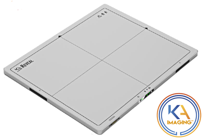
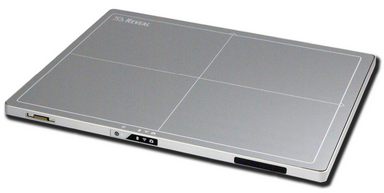
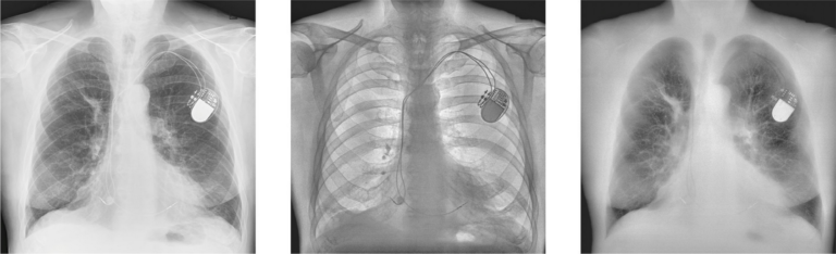
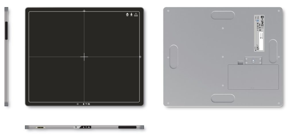
組裝
我們是醫療器材OEM、ODM 製造商。提供完整的設計和製造的服務.。
同時,我們也生產製造 X光數位平板感測器的關鍵零組件與配件。
Reveal™ is the world’s first mobile digital detector capable of separating different energy levels in a single exposure. Without motion artifacts, it simultaneously captures dual-energy images and very-high DQE digital radiography (DR) images. Using KA Imaging’s patented technology, Reveal differentiates bone and soft tissue while maintaining low exposure to radiation.
The future is now and cost-effective
Reveal™ is designed to obtain a Multi-Energy X-Ray image from a single exposure using a conventional X-Ray source. The patented technology features a portable and retrofittable design, capable of simultaneously capturing Multi-Energy images and very high DQE digital radiography (DR) images.
- Soft-tissue differentiation
- Bone and soft tissue differentiation
- Single exposure, no motion artifact
- Retrofittable
- Fixed and mobile applications
- 80% DQE
- Portable and retrofittable
Flexible applications
Reveal™ features a portable design, which makes it ideal for fixed and mobile DR
applications. The detector has been designed in the standard cassette size, allowing for retrofit in any existing x-ray system.
In medical applications, dual-energy technology has been clinically proven to be effective
in imaging and diagnosing pulmonary disease
Globally, lung cancer is the leading cause of cancer deaths, numbering over 150,000 deaths each year in the US alone. Despite the high incidence and death rate with this type of cancer, screening options remain limited and costly to implement for high-risk individuals. Often, lung cancer is diagnosed in late stages of the disease, limiting treatment options and lowering the rate of survival. Currently, the most commonly used method of screening on the market is Low-Dose Computed Tomography. Though effective, this method suffers from low availability and still exposes patients to high doses of radiation, which results in low follow-up numbers.
Clinical Trial
In October 2018, KA Imaging initiated a clinical trial at Grand River Hospital AL in Kitchener, Ontario, Canada to image lung cancer patients with Reveal™. TRIAL In this trial, more than 20 patients have been scanned using the detector to visualize lung nodules and lesions, with promising initial results. Chest X-Ray images displayed are from the clinical trial.
Smart design for Hot swap and seizing image in time
Access point mode
Equipped with access point function, our product can connect to computer without extra wireless configuration setting. It undoubtedly brings a user-friendly interface to improving work efficiency.
Overview
Specification
Detector Size (ISO 4090) | 14 x 17 inch / 35 x 43 cm |
Pixel Pitch | 140 um |
Sensor Type | Amorphous Silicon |
Scintillator | Csl |
DQE (0) | 80% for RQA5 |
Communication | Wireless or gigabit ethernet |
Trigger | Software and lossless AED available |
Power | Battery and/or tethered AC 100-240 VAC/50-60 Hz |
A/D Conversion | 16 bits |
Cycle Time | 7 seconds |
Preview Latency | Typical time 4 seconds for wireless mode |
Software | C++ SDK available for system integration |
Energy Range | 40-150 kVp* |